
Figure 3. Increased STAT3 activation in tongue epithelium of infected mice.
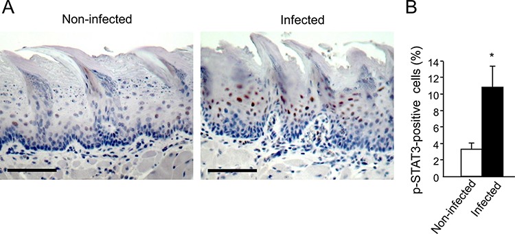
C57/Bl6 mice were infected with a mixture of P. gingivalis and F. nucleatum (as described in Methods) every other day for 6 days. Mice were sacrificed twenty-four hours after the last infection, their tongues removed and processed for immunostaining with anti-pSTAT3 antibody. A. Immunostaining with anti pSTAT3 antibody reveals increased levels of nuclear-localized pSTAT3 in tongue epithelium of infected (right) vs. non-infected (left) mice. Representative images of are shown. Magnification × 200 B. Percentage of pSTAT3-immunostained cells in the epithelium was calculated in ≥5 microscopic fields per mouse. Data are the mean ± SE, *p = 0.01 (Student's t-test). Scale bars: 100 μm.