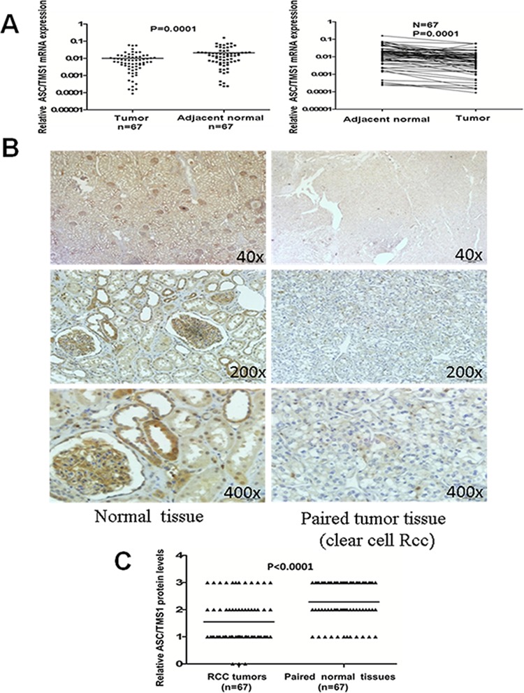
Figure 2. Expression pattern of ASC/TMS1 in RCC.

A. The mRNA expression levels of ASC/TMS1 in paired primary RCC tissues as determined by quantitative real-time PCR. ASC/TMS1 mRNA was significantly downregulated in RCC samples compared with their adjacent normal tissues (p = 0.0001). B. Representative immunohistochemical staining of a pair of RCC specimens and corresponding nontumor tissues. In adjacent nontumor tissues, intense immunostaining for ASC/TMS1 was detected in a cytoplasmic and nuclear distribution, whereas absent/weak immunostaining was observed in the cytoplasm and nucleus of tumor tissues. C. Evaluation and statistical analysis of ASC/TMS1 protein expression in 67 paired primary RCC tissues. ASC/TMS1 protein expression was significantly downregulated in RCC samples compared with adjacent normal tissues (P < 0.0001).
