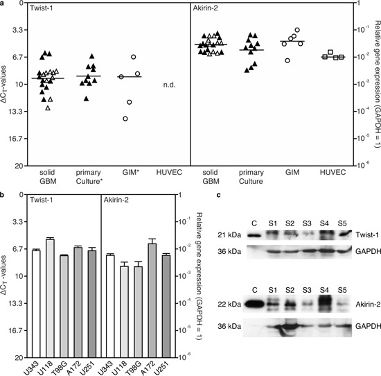
Figure 1. Expression of Akirin-2 and Twist-1 in a. solid and matched cultured primary human glioblastomas (GBM), glioma-infiltrating macrophages/microglia (GIM), human umbilical vein endothelial cells (HUVEC) and b. GBM cell lines was evaluated by real-time RT-PCR (measured in duplicates; logarithmic scale, ΔCT = 3.33 corresponds to a 10-fold difference; black filled symbols identify matched samples of solid and cultured GBMs), and c. Western Blot analysis of Akirin-2 and Twist-1 expression in five different solid (s) GBMs compared to recombinant Akirin-2 or Twist-1 control (c) proteins.

a, b. Both Akirin-2 and Twist-1 were found in distinct mRNA expression amounts in all investigated samples with Akirin-2 in comparable high level. An asterisk (*) symbolizes one Twist-1 value below detection limit in primary cultures or GIMs, respectively. c. Both Akirin-2 and Twist-1 proteins were detectable with varying amounts in different human GBM samples yielding expected sizes of 22 kDa and 21 kDa, respectively. To confirm protein integrity, blots were reprobed with glycerinaldehyde-3-phosphate-dehydrogenase (GAPDH). Representative examples of three independent experiments are shown.
