
Figure 1. Acadesine induces apoptosis and activates AMPK.
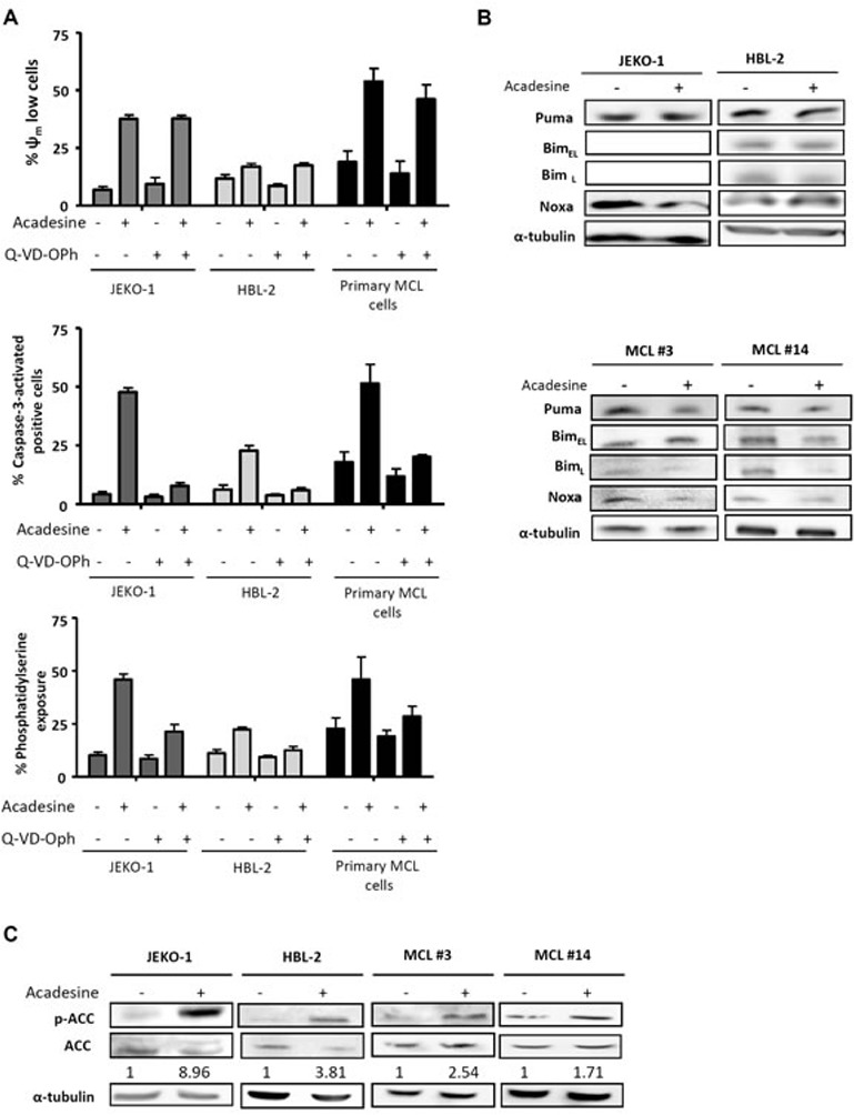
A. JEKO-1, HBL-2 and 3 primary MCL samples were preincubated for 1 hour with 10 μM of the pan caspase inhibitor Q-VD-OPh and followed by a 24-hour exposure to acadesine 2 mM. Mitochondrial membrane potential (Ψm), caspase-3 activation and phosphatidylserine exposure were evaluated by flow cytometry as detailed in “Methods”. B. MCL lines (JEKO-1 and HBL-2) and two representative primary MCL samples were cultured with acadesine 2 mM for 6 hours and protein levels of Bim, Puma and Noxa were determined by western blot. α-tubulin was used as loading control. C. MCL lines (JEKO-1 and HBL-2) and two MCL primary samples were cultured with acadesine 2 mM for 6 hours. Phosphorylated and total levels of ACC were assessed by western blot using α-tubulin as loading control. The ratio between the phosphorylated and unphosphorylated form was showed.