
Figure 3. Acadesine downregulates Mcl-1 through the mTOR pathway.
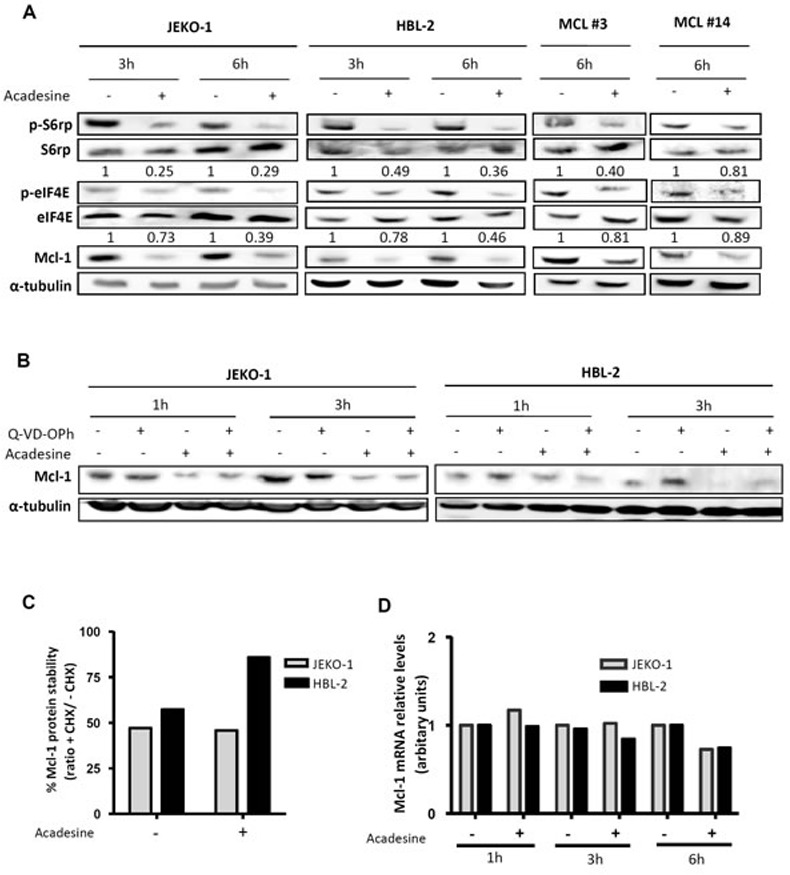
A. MCL lines (JEKO-1 and HBL-2) and two primary MCL samples were incubated with acadesine 2 mM for the indicated times. Phosphorylation of kinases of the AMPK/mTOR pathway and Mcl-1 levels were determined by western blot using α-tubulin as a loading control. The ratio between the phosphorylated and unphosphorylated form was showed. B. JEKO-1 and HBL-2 cells were cultured with acadesine 2 mM after a 1-hour incubation with Q-VD-OPh for the indicated times. Mcl-1 levels were detected by western blot using α-tubulin as loading control. C. MCL cells (JEKO-1 and HBL-2) were treated for 1 hour with cycloheximide prior to incubation with acadesine 2 mM for 4 hours. Relative levels of Mcl-1 were quantified by densitometry using α-tubulin as a loading control. Bars represent the protein stability considering the ratio with/without cycloheximide. CHX, cycloheximide. D. Relative Mcl-1 mRNA levels were quantified in JEKO-1 and HBL-2 after acadesine (2 mM) exposure for the indicated times by qRT-PCR taking as a reference the corresponding untreated condition.