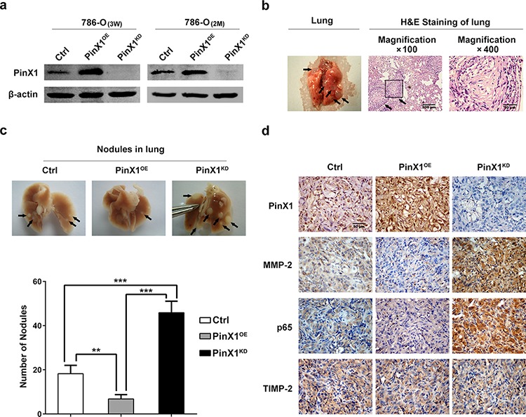
Figure 5. PinX1 suppress ccRCC metastasis in vivo.

a. left panel, Western blotting of PinX1 from PinX1OE-786-O cell lines, PinX1KD-786-O cell lines and Ctrl-786-O cell lines selected with puromycin for 3 weeks after lentivirus infection. Right panel, PinX1 expression levels were not changed in PinX1OE, PinX1KD and Ctrl 786-O cell lines without puromycin selection for 2 months. b. Representative image of lung with metastatic nodules (left panel) and H&E staining sections of lung (right panel) 2 months after injection of PinX1KD 786-O cell lines in BALB/c nude mouse through tail vein. Arrows indicate metastatic nodules. c. top panel, Representative images of 10% buffered formalin fixed lungs with metastatic nodules 2 months after respective injection of Ctrl, PinX1OE and PinX1KD 786-O cell lines. Arrows indicate metastatic nodules. Bottom panel, the number of lung metastatic nodules was counted under a dissecting microscope. A statistically dramatic increase in the number of the lung metastases was seen in PinX1KD group, compared with the PinX1OE group and these two groups also had significant diversity compared with Ctrl group respectively. Data are displayed with means ± SD from 12 mice in each group. **, P < 0.01; ***, P < 0.001. d. Immunostaining of PinX1, MMP-2, p65 and TIMP-2 in metastatic nodules of PinX1OE, PinX1KD and Ctrl 786-O groups. MMP-2 and p65 expression in PinX1OE group were much lower compared with PinX1KD group and Ctrl group but TIMP-2 expression was not change in every group.
