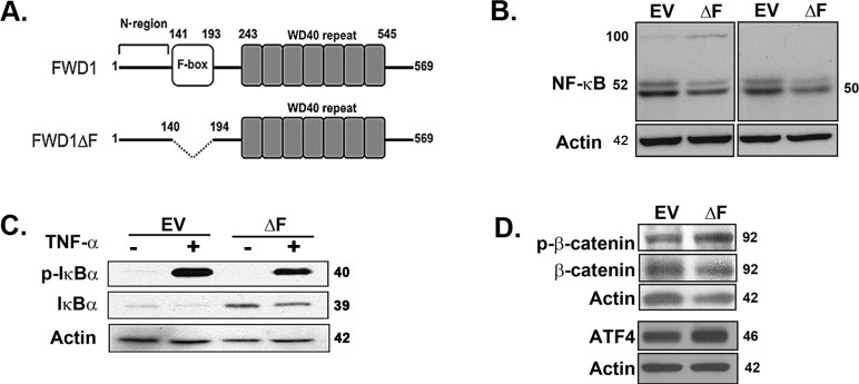
Figure 1. Dominant-negative expression of β-TrCP1/FWD1 disrupts NF-κB signaling in myeloma cells.
A. Representation of β-TrCP/FWD1ΔF (adapted from [12]). B. Immunoblotting for p50/105 (left panel) and p52/p100 (right panel) in lysates obtained from untreated 5TGM1-ΔF and 5TGM1-EV cells shows increased accumulation of p100 and decreased p52 levels in 5TGM1-ΔF myeloma cells. C. Cells were treated with 20 ng/ml TNF-α and lysates prepared. Immunoblotting shows increased basal levels of total IκBα in 5TGM1-ΔF cells. D. Untreated 5TGM1-ΔF and 5TGM1-EV cells were probed for ATF4, and for total and phosphorylated forms of β-catenin. Blots were normalized to actin.

