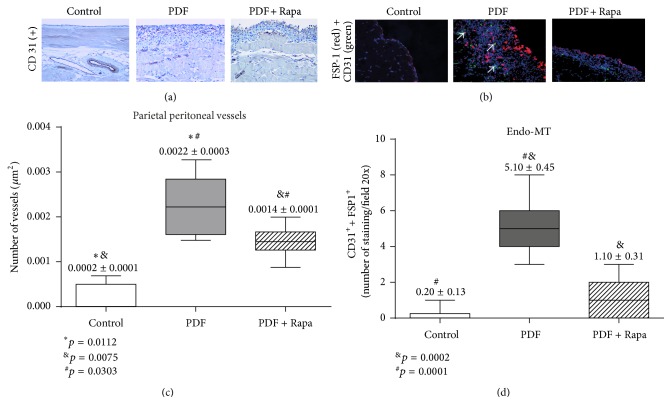
Figure 2.
Treatment with Rapamycin decreases PD-induced angiogenesis and Endo-MT. Mice received a daily instillation of standard PD fluid with or without the oral administration of Rapamycin (PDF; n = 5 and PDF + Rapamycin; n = 6). A control group of mice was also included (control; n = 6). (a) Standard PD fluid exposure increases peritoneal angiogenesis and Rapamycin administration significantly reduces the number of submesothelial blood vessels, as determined by CD31 staining (number of CD31+ cells/μm2). Magnification 200x. (b) Standard PD fluid exposure also increases the presence of endo-MMT, measured as double positive immunofluorescence staining (white arrows) of CD31 (green) and FSP-1 (red) (counterstained with DAPI, in blue), which is reduced by Rapamycin administration. Magnification 200x. (c) Box plots represent the number of submesothelial CD31+ vessels stained cells per field in the different experimental groups and show a decrease of angiogenesis in the Rapamycin-treated animals. The analysis of variance results in a significance of p < 0.0001 (one-way ANOVA test). (d) The numbers of double stained CD31+/FSP-1+ cells per field increase in the PDF-exposed group and show a decrease in the Rapamycin-treated animals. The one-way ANOVA test resulted in a significance of p < 0.0001. Box plots graphics represent 25th and 75th percentiles and median, minimum, and maximum values. Numbers above boxes depict means ± SE. Symbols represent the statistic differences between groups.

