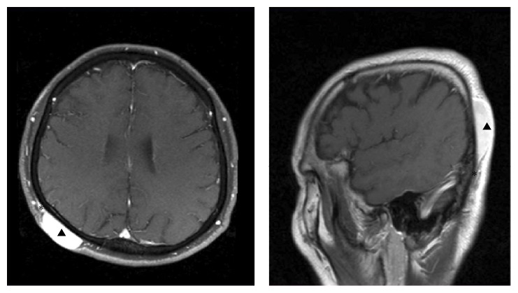
Figure 2.
MRI image showing the subcutaneous extension of DFSP. These images help to determine the outline of DFSP. Arrow heads present primary lesion of DFSP in scalp. Asterisks present subcutaneous extension beyond the macroscopic tumor margin. MRI: magnetic resonance imaging, DFSP: dermatofibrosarcoma protuberans.

