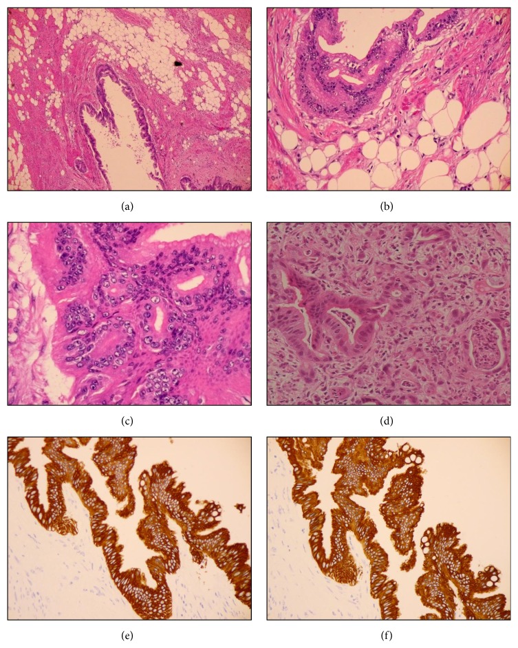
Figure 4.
((a)–(c)) Photomicroscopy images showing lipoleiomyoma infiltrated with well- to moderately differentiated adenocarcinoma. (d) Biopsy specimen from the previous endoscopy showing poorly-differentiated adenocarcinoma. Stain, hematoxylin and eosin. ((e) and (f)) Representative immunohistochemistry for (e) CK7 and (f) CK20. Both CK7 and CK20 were positive in well- to moderately differentiated adenocarcinoma.

