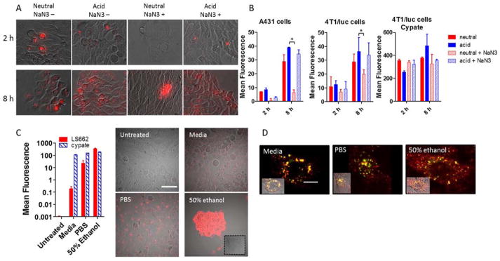
Figure 3.
(A) Images of cellular uptake of LS662 in A431 cells for 2 or 8 h at normal physiological pH 7.4 (neutral) or acidic pH 6.4 (acid) media. Sodium azide (NaN3+) was added to each condition to determine whether the internalization of LS662 is energy dependent. (B) Quantification of the cellular uptake, described in A, in A431 and 4T1/luc cells with LS662 and cypate, *p-value < 0.05. (C) Cellular uptake of LS662 and cypate in A431 cells treated in three conditions: media, PBS, and 50% ethanol, to probe healthy, dying and dead cells, respectively. The images show LS662 fluorescence; cypate images not shown. (D) Healthy, dying and dead cells were coincubated with LS662 (red) lysotracker (green), to investigate the subcellular localization of LS662. Scale bar = 10 μm.

