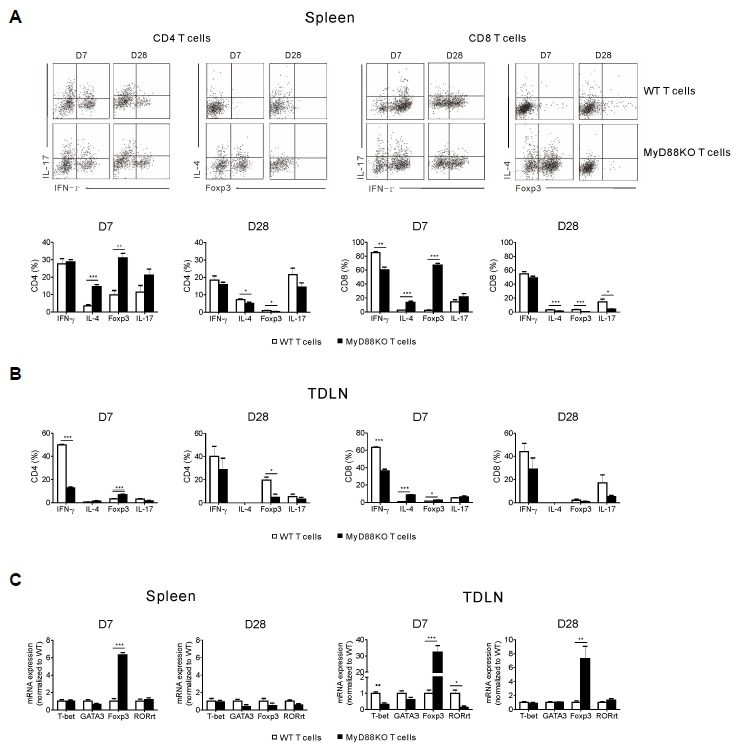
Fig. 4.

Absence of MyD88 in donor T cells leads to an expansion of Treg and TH2 cells with reduced TH1 cells in lymphoid organs. Lethally irradiated recipient mice were transplanted with WT TCD-BM (5 × 10⁶) together with either WT or MyD88KO mice spleen T cells (1 × 10⁶) on day 0. All animals (n = 6, each group) were also injected s.c. with 1 × 10⁶ P815 tumor cells on day 1. Spleens (A) and tumor-draining lymph node (TDLN) (B) were harvested from the recipient mice on days 7 and 28 post-transplantation. Expressions of IFN-γ, IL-4, Foxp3 and IL-17 on the CD4+ and CD8+ T cells were assayed by FACS. (C) Expression of T-bet, GATA3, Foxp3 and RORγT were measured in the mononuclear cells of spleen and TDLN using quantitative RT-PCR. Data are presented as means ± SEMs and are representative of duplicate experiments (*P < 0.05, **P < 0.01 or ***P < 0.001).
