
Fig. 2.
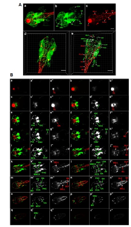
SPIM and 3D reconstruction of cranial neurons and vasculature in Tg(elavl3:DsRed/ fli1a: EGFP) zebrafish embryo at 42 hpf. (A) Dorsal view of whole head and anterior trunk. (a–c) 3D reconstruction of the entire anterior portion of embryo. fli1a:EGFP-positive cells are green and elavl3:DsRed-positive cells are red. Data is comprised of 479 optical sections of 1.176 μm thickness using optimum interval option. Scale bar 100 μm. An animated movie of the 3D raw data analysis is available in Supplementary movie Fig. S2A. (d, e) 3D reconstruction of selected optical sections (277–362) (d) and its 3D segmentation (e) using Imaris software to reveal fine detail of internal structure. Scale bar 150 μm. Video files of the final 3D reconstructions are presented as Supplementary movies in Figs. S2B (raw data) and S2C (segmented images). (B) Optical sections of 42 hpf embryo. Optical sections covering the entire embryonic head along the dorsalventral axis were merged into 21 consecutive slices of 24.696 μm thickness. The 18 of these sections (a–r) are shown (a′–r′:fli1a: EGFP, a″–r″: elavl3:DsRed). BA, basilar artery; BCA, basal communicating artery; CA, caudal artery; CCA, cerebellar central artery; CCV, common cardinal vein; CMV, communicating vessel; CVP, choroidal vascular plexus; DA, dorsal aorta; DELL, diencephalic efferent neurons to the lateral line; DLF, dorsal longitudinal fasciculus; DMJ, dorsal midline junction; IOC, inner optic circle; LDA, lateral dorsal aorta; MMCA, middle mesencephalic central artery; MsA, mesencephalic artery; MV, mesencephalic vein; NC, notochord; NCA, nasal ciliary artery; ON, optic neuron; OV, otic vesicle; PA, pharyngeal arch; PCS, posterior communicating segment; PF, pectoral fin; PHC, primordial hindbrain channel; PLLG, posterior lateral line ganglion; PrA, prosencephalic neuron; RB, Rohon-Beard neuron; SIV, subintestinal vein; SN, spinal neuron; TC, telencephalic cluster, 1, mandibular arch; 2, hyoid arch; 3 to 7, pharyngeal arch.