
Fig. 3.
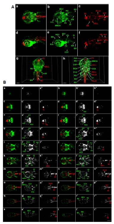
3D imaging analysis of cranial neurons and vasculature in Tg(elavl3: DsRed/Fli1a:EGFP) zebrafish embryo at 3.2 dpf. (A) Dorsal view of whole head and anterior trunk. (a–c) 3D reconstruction: fli1a:EGFP-positive tissues are green and elavl3:DsRed-positive neurons are red. Data set is 503 μm thick. Scale bar 200 μm. (d–f) Lateral images of the same embryo: 591 μm thickness. (g) 3D reconstruction of raw data: 503 μm thickness. (h) 3D segmentation of selected sections: 277 μm thickness, internal structures are labelled. Scale bars 150 μm. 3D movies showing the entire reconstructed head are presented in Supplementary Figs. S3A (raw data) and S3B (segmented images). (B) Optical sections of Tg(elavl3:DsRed/fli1a:EGFP) zebrafish at 3.2 dpf (a–t). Optical sections were combined to create 21 μm compressed sections highlighting fli1a:EGFP-positive tissues (green; a′–t′) and elavl3:DsRed-positive neurons (red; a″–t″). AMCA, anterior mesencephalic central artery; BA, basilar artery; BCA, basal communicating artery; CaDI, caudal division of the internal carotid artery; CCA, cerebellar central artery; CCV, common cardinal vein; CrDI, cranial division of the internal carotid artery; CVP, choroidal vascular plexus; DELL, diencephalic efferent neurons to the lateral line; DLF, dorsal longitudinal fasciculus; DLV, dorsal longitudinal vein; DMJ, dorsal midline junction; IOC, inner optic circle; IV, intersegmental vessel; MCV, middle cerebral vein; MMCA, middle mesencephalic central artery; MV, mesencephalic vein; NC, notochord; NCA, nasal ciliary artery; ON, optic neuron; PA, pharyngeal arch; PCS, posterior communicating segment; PF, pectoral fin; PG, pronephric glomus; PHC, primordial hindbrain channel; PICA, primitive internal carotid artery; PLA, palatocerebral artery; PLLG, posterior lateral line ganglion; PLLN, posterior lateral line neuron; PrA, prosencephalic neuron; RB, Rohon-Beard neuron; RELL, rhombencephalic efferent neurons to the lateral line; SIA, subintestinal artery; SIV, subintestinal vein; SN, spinal neuron; TC, telencephalic cluster; 1, mandibular arch; 2, hyoid arch; 3 to 7, pharyngeal arch.