
Fig. 4.
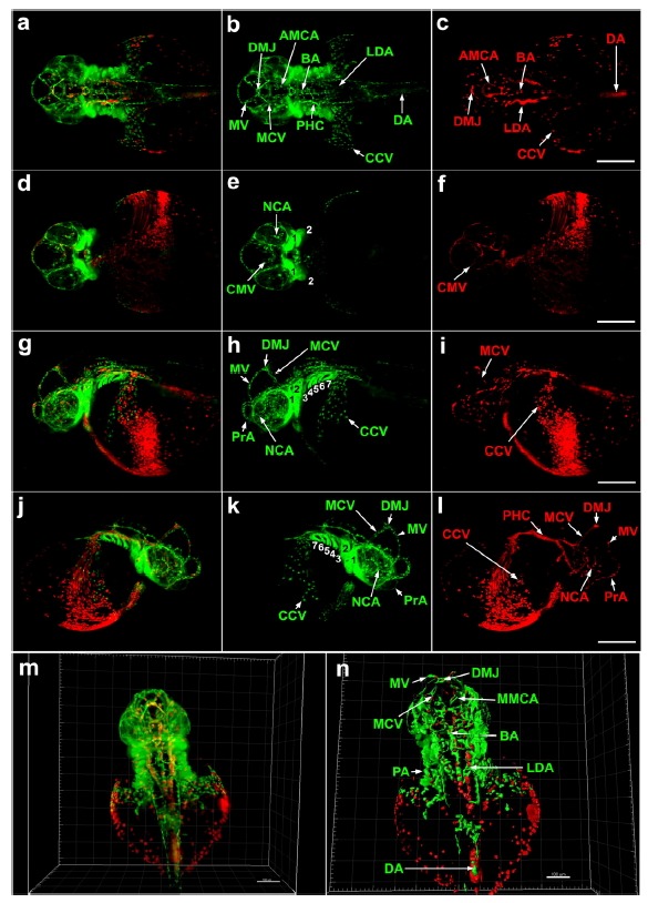
Multi-view SPIM imaging of embryonic blood cells and vasculature in Tg(gata1: DsRed/fli1a:EGFP) zebrafish at 35 hpf. (a–l) 3D reconstruction from 1.0 μm optical sections of whole brain and anterior trunk (including subintestinal vasculature) of 35 hpf embryo. fli1a: EGFP-positive cells are green gata1:DsRed -positive blood cells are red. (a–c) Dorsal view, constructed from 399 optical sections (280–618). (d–f) ventral view, constructed from 457 optical sections (195–651). (g–i) left lateral view: 515 optical sections (225–739). (j–l) right lateral view: 445 optical sections (292–736). Scale bars 200 μm. (m) 3D reconstruction of raw data containing 410 optical sections (280–618) (n) 3D reconstruction of segmented data using Imaris software’s surface function. Scale bar, 150 μm. Supplementary Figs. S4A and S4B show final 3D reconstructions from raw and segmented data. AMCA, anterior mesencephalic central artery; BA, basilar artery; CCV, common cardinal vein; CMV, communicating vessel; DA, dorsal aorta; DMJ, dorsal midline junction; LDA, lateral dorsal aorta; MCV. middle cerebral vein; MMCA, middle mesencephalic central artery; MV, mesencephalic vein; NCA, nasal ciliary artery; PHC, primordial hindbrain channel; PrA, prosencephalic artery; 1, mandibular arch; 2, hyoid arch; 3 to 7, pharyngeal arches.