Figure 3.
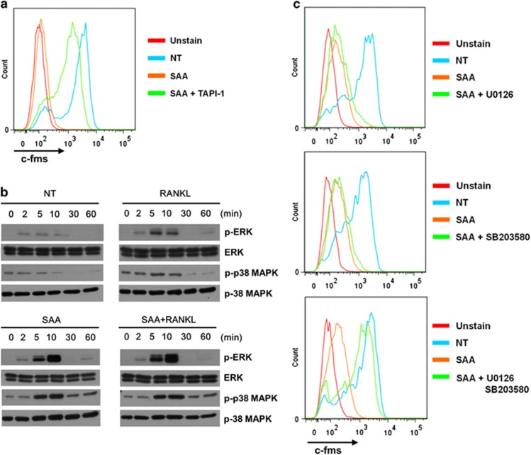
SAA stimulates c-fms shedding by TACE, which is dependent on ERK and p38 MAPK activity. (a) Mouse BMDMs were pre-incubated with TAPI-1 (20 μM) for 1 h prior to the SAA (1 μM) treatment. The c-fms levels on the cell surface were determined by flow cytometry using an anti-c-fms antibody. (b) Mouse BMDMs were stimulated with SAA (1 μM) in the presence of M-CSF (30 ng ml−1), RANKL (100 ng ml−1) or M-CSF (30 ng ml−1) and RANKL (100 ng ml−1) for 0, 2, 5, 10, 30 and 60 min. The phosphorylated ERK and p38 MAPK levels were determined by immunoblotting using anti-phospho-ERK and anti-phospho-p38 MAPK antibodies. (c) Mouse BMDMs were pre-incubated for 1 h with U0126 (40 μM; top), SB203580 (20 μM; middle), or U0126 (40 μM) and SB203580 (20 μM; bottom) prior to the SAA (1 μM) treatment. The c-fms levels on the cell surface were determined by flow cytometry using an anti-c-fms antibody. The results shown are representative of three independent experiments (a–c). BMDM, bone marrow-derived macrophage; M-CSF, macrophage colony-stimulating factor; NT, not treated; RANKL, receptor activator of nuclear factor κB ligand; SAA, serum amyloid A.

