Figure 1.
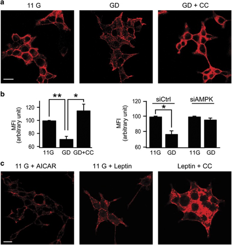
Glucose deprivation or leptin induces actin disruption via AMPK signaling in INS-1 cells. (a, c) Confocal fluorescent images of INS-1 cells stained with Alexa 633-phalloidin for F-actin staining. (a) Before fixation, cells were pretreated with 11 mM glucose (11 G) or 0 mM glucose for 2 h in the absence (GD) or presence of 10 μM compound C (GD+CC). Scale bars, 10 μm. (b) Quantification of changes in F-actin content using FACS analysis. Mean fluorescence intensity (MFI) of 11G was arbitrarily set at 100% and compared with MFI of GD and GD+CC cells (left). MFI of siAMPK-transfected GD cells is compared with siCtrl-transfected GD cells (right). Data are represented as the mean±s.e.m. of three independent experiments. *P<0.05 and **P<0.01. (c) Cells were incubated in 11 mM glucose for 30 min in the presence of 0.25 mM AICAR (11 G+AICAR) or 10 nM leptin in the absence (11 G+Leptin) and the presence of 10 μM CC (Leptin+CC). Scale bars, 10 μm.

