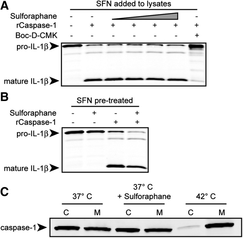
Figure 3. SFN does not inhibit recombinant caspase-1 (rCaspase-1) directly, induce a cellular state inhibitory to rCaspase-1 activity, or sequester procaspase-1 into a high-molecular-weight complex.
(A) RAW264.7 (pictured) or Balb/cJ BMDM cells were treated with LPS (1 μg/ml) for 2 h, and sucrose lysates were prepared. Boc-D-CMK (400 μM) or SFN at varying concentrations (0, 6.25, 12.5, 25, or 50 μM) was added to cell lysates. (B) RAW264.7 (pictured) or Balb/cJ BMDM cells were treated with LPS (1 μg/ml) for 2 h, then with SFN (50 μM) for 1 h before preparation of sucrose lysates. (A and B) Murine active recombinant caspase-1 (rCaspase-1) was added to the lysates. Lysates were incubated at 37°C for 3 h. To measure rCaspase-1 activity, IL-1β processing was evaluated by Western blot. (C) Balb/cJ BMDMs were pretreated with or without SFN (50 μM) at 37°C or without SFN at 42°C for 1 h. Sucrose lysates were prepared and fractionated by centrifugation into cytosolic (C) or membranous (M) fractions. Western blotting for caspase-1 was performed to determine its localization to either the cytosol or membrane-bound, high-molecular-weight protein complex. All data are representative of 2 or more independent experiments.

