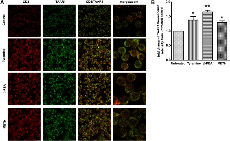
Figure 2. METH increases TAAR1 protein expression in resting human T cells.
(A) Immunofluorescent staining and confocal imaging of human T lymphocytes showing expression of TAAR1. Representative photomicrographs of CD3 (red) and TAAR1 (green). One million T lymphocytes were left untreated or incubated for 24 h at 37°C with 100 μM of the one following treatments: β-PEA (Sigma-Aldrich), tyramine (Sigma-Aldrich), or MET (Sigma-Aldrich). (B) Data represent the fold change of the average of mean gray-pixel values ± sem from 5 separate images/treatment were taken at 63×. Images were analyzed for mean gray pixels/cells in the field by use of ImageJ software. T cells treated with tyramine (100 μM), β-PEA (100 μM), and METH (100 μM) showed a significant increase in TAAR1 expression compared with untreated control cells. *P < 0.05 for METH and tyramine; **P < 0.01 for β-PEA.

