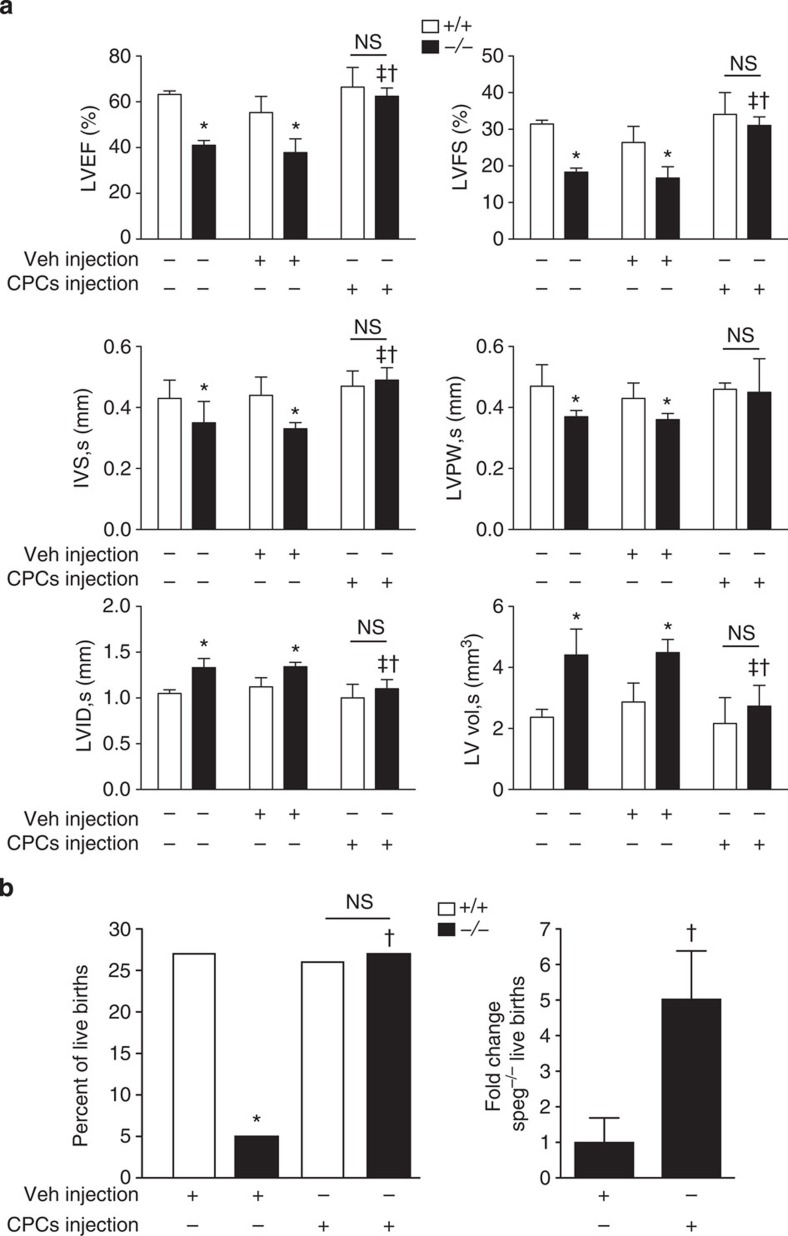
Figure 10. Functional assessment of Speg−/− and Speg+/+ hearts after in utero injections of CPCs.
(a) Speg+/+ (white bars, n=6–7 per group) and Speg−/− (black bars, n=4–5 per group) mice received either no injection (−), or intra-cardiac injection (+) with vehicle (Veh) or CPCs at 13.5 dpc. On day 1 after birth (19.5 dpc), echocardiograms were performed to assess cardiac function. The left ventricles were assessed for ejection fraction (LVEF) and fractional shortening (LVFS). Measurements for thickness of the intraventricular septum (IVS) and LV posterior wall (LVPW), and dimensions of the LV internal diameter (LVID) and LV volume (LV vol) were performed during systole (s). Data are presented as mean±s.e.m. *P<0.05 versus Speg+/+ mice in the same group. ‡P<0.05 versus Speg−/− mice receiving no injection. †P<0.05 versus Speg−/− mice receiving Vehicle injection. NS, not significant. Analyses performed using one-way analysis of variance, followed by either Bonferroni's or Newman–Keuls multiple comparison test. (b) Foetuses of Speg+/− pregnant dams were injected with either Vehicle (Veh, n=6 litters) or wild-type CPCs (n=6 litters) at 13.5 dpc. On day 1 after birth (19.5 dpc), the per cent of live births was assessed in Speg+/+ (white bars) and Speg−/− (black bars) pups (left panel). P=0.0012; * versus Speg+/+ pups, Vehicle injection; † versus Speg−/− pups, Vehicle injection. Analysis performed by Fisher's exact test. In the right panel, fold change (mean±s.e.m.) in live births of Speg−/− pups receiving either Vehicle or wild-type CPCs injection. P=0.0198; † versus Speg−/− pups, Vehicle injection using Student's unpaired t-test.

