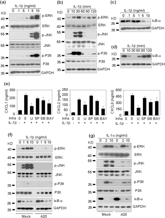
Figure 9. The signal transduction for CXC chemokine production induced by IL-1.
(a) IL-1β dose dependent activation of MAPKs. HMC cells were treated with the indicated concentrations of IL-1β for 30 min. The phosphorylation of MAPKs ERK (42/44 KD), JNK 46/54 KD), and p38 (38 KD) was detected by western blot. GAPDH (36 KD) protein levels were detected as loading controls. (b) IL-1β time-dependent activation of MAPKs. HMC cells were treated with 10 ng/ml IL-1β for the indicated time periods. The phosphorylation of MAPKs ERK, JNK, and p38 was detected by western blot. GAPDH protein levels were detected as loading controls. (c) IL-1β dose-dependent degradation of IκB-α (39 KD). (d) IL-1β time-dependent degadation of IκB-α. (e) The effect of signal inhibitors on chemokine induction. HMC cells, pre-treated without medium, or ERK inhibitor U0126 (U, 10 μM), or JNK inhibitor SP600125 (SP, 10 μM), or P38 inhibitor SB203580 (SB, 10 μM), or NF-κB inhibitor Bay 117082 (Bay, 10 μM) for 30 min, were re-stimulated with 5 ng/ml IL-1β for 6 h. Chemokine protein levels in the culture supernatant were detected by ELISA. p-value *< 0.05 compared with the IL-1β-treated alone groups. (f) The effect of A20 over-expression on IL-1β-induced signal transduction. Mock- and A20-transfected HMC cells were treated with the indicated concentration of IL-1β for 30 min. The phosphorylation of MAPKs, and the total protein levels of IκB-α were detected as same as (a). (g) The effect of A20 over-expression on IL-1α-induced signal transduction. p-value *< 0.05 compared with the IL-1β-treated alone groups.

