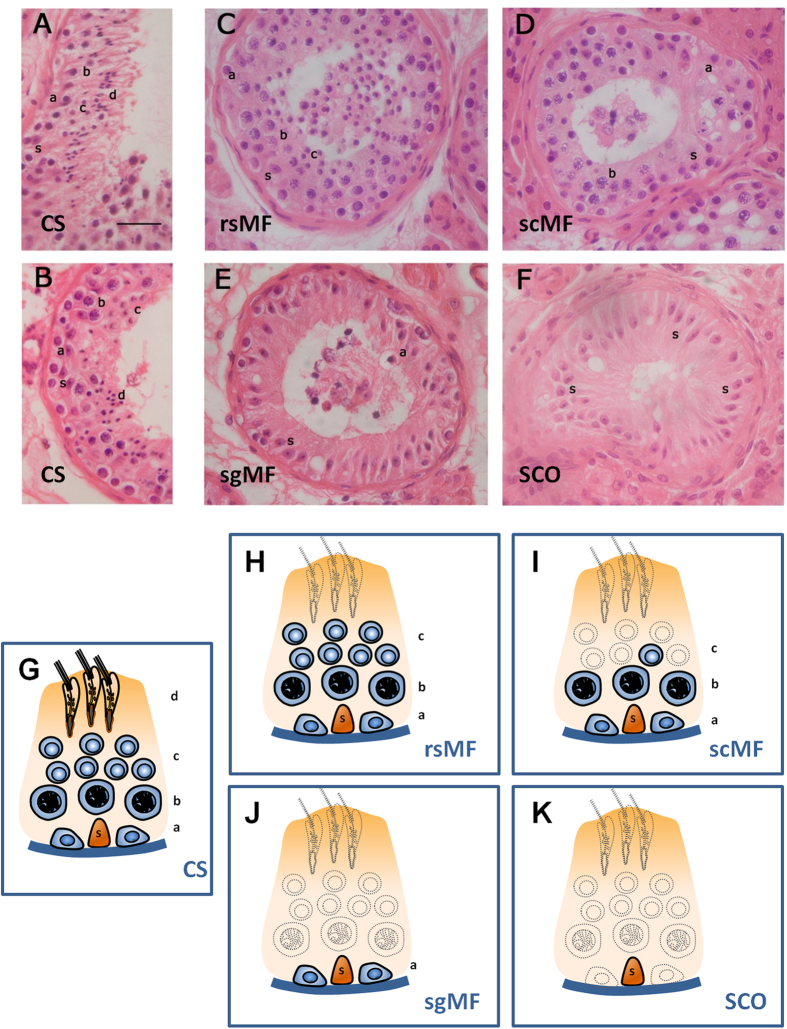
Figure 1. Testicular histology of representative sections of seminiferous tubules from infertile men showing different spermatogenic patterns: conserved spermatogenesis (CS) containing all germ-cell stages (A,B), maturation failure at the round spermatid stage (rsMF) showing no elongated spermatids (C), maturation failure at the spermatocyte stage (scMF) with spermatogonia and spermatocytes but few or none spermatids (F), maturation failure at the spermatogonia stage (sgMF) displaying only these primordial cells (G), and Sertoli cell-only phenotype or germ-cell aplasia (SCO) where seminiferous tubules contain exclusively Sertoli cells (H).
Bellow, schematic diagrams, showing the epithelium of a seminiferous tubule that consists of a single Sertoli cell and the germ-cell components of the different phenotypes, are placed below the histological images: CS (G), rsMF (H), scMF (I), sgMF (J), and SCO (K). Absent cell populations are drawn as dashed line shadings. Examples of different cell types, including Sertoli cells and specific germ-cell stages, are identified with lower case letters: (a): spermatogonia; (b): primary spermatocytes; (c): round spermatids; (d): elongated spermatids; (s): Sertoli cell nucleus. Hematoxilin and eosin staining. Bar = 50μm.

