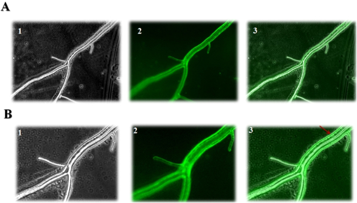
Figure 2. Fluorescence microscopy analyses of Epl-1 fused with GFP.
(A) – Fluorescence microscopy of Trichoderma harzianum RecEpl-1-GFP hyphae in 20× optical magnification. 1 – Light field; 2 – GFP; 3 – Merge. (B) – Fluorescence microscopy of Trichoderma harzianum RecEpl-1-GFP strain hyphae in 40× optical magnification. 1 – Light field; 2 – GFP; 3 – Merge. The arrow indicates the predominance of Epl-1 in the cell wall region and its transport through the hyphae by vesicles.

