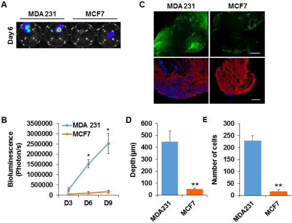
Figure 3. Metastatic MDA-MB 231 cells grow and invade in the decellularized lung matrix.
(A) IVIS Images showed the growth of MDA-MB 231-Luc cells and MCF7-Luc cells in decellularized lung tissue on Day 6. (B) Quantification of the IVIS results showed the number of MDA-MB 231-Luc cells and MCF7-Luc cells on Day 3, Day 6 and Day 9; n=4. *, p<0.05 (C) Upper panel: Fluorescence images showed the growth of GFP-labeled MDA-MB 231 and MCF7 cells in decellularized lung on Day 9. Bottom panel: IF staining of ColIV (red) and DAPI (Blue) staining on sections of decellularized tissue cultured with MDA-MB 231-Green cells and MCF7-Green cells. Scale bar: 200 μm. (D, E) Bar graphs of the invaded depth (D) and number (E) of MDA-MB 231-Green cells and MCF7-Green cells in the decellularized lung tissue; n=5. **, p<0.01.

