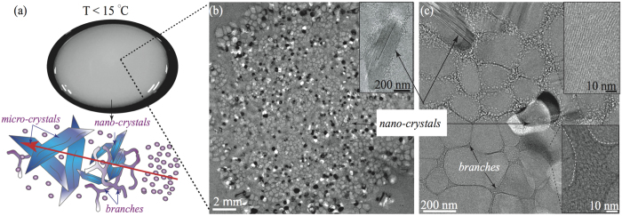
Figure 4.
(a) The CLS exhibits turbid and “milky” appearance. The cartoon represents the transition from spherical micelles to CLS. Different sizes of needle-like blue objects represent either nanocrystal-like or microcrystal-like entities. (b) Low magnification cryo-EM image of the CLS: showing wormlike micelles, branched networks, and nano-crystals (white-black domains and inset). (c) High magnification cryo-EM images of the CLS, exhibiting wormlike micelles and branched networks. CLS consists of branched micellar bundles, formed from individual wormlike micelles. The insets display the stacking of individual wormlike micelles. Wormlike micelles in the CLS have a diameter of ~9.4 ± 0.3 nm, while the micellar branches diameter ranges from tens to hundreds of nanometers.

