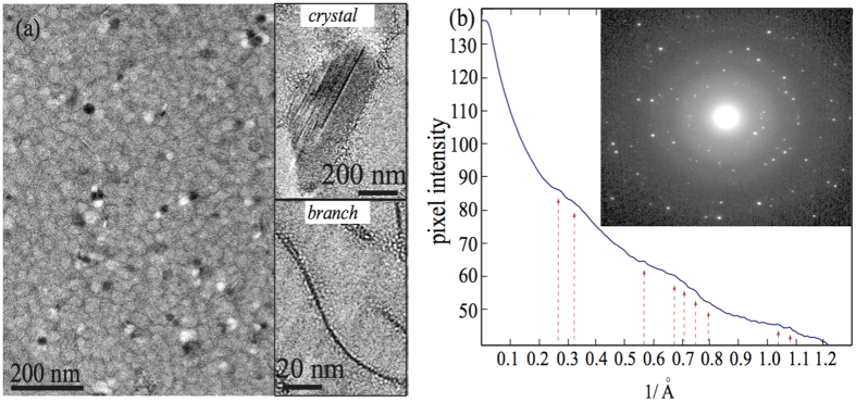
Figure 6.
(a) Representative cryo-EM image of the CLS. The white and black spots are nanocrystal-like domains in the CLS. The insets display the crystal-like structures and micellar branches in the CLS. (b) Rotationally averaged radial intensity obtained from the CLS. The inset shows the diffraction patterns of the CLS, exhibiting some symmetry and lattice order in the CLS.

