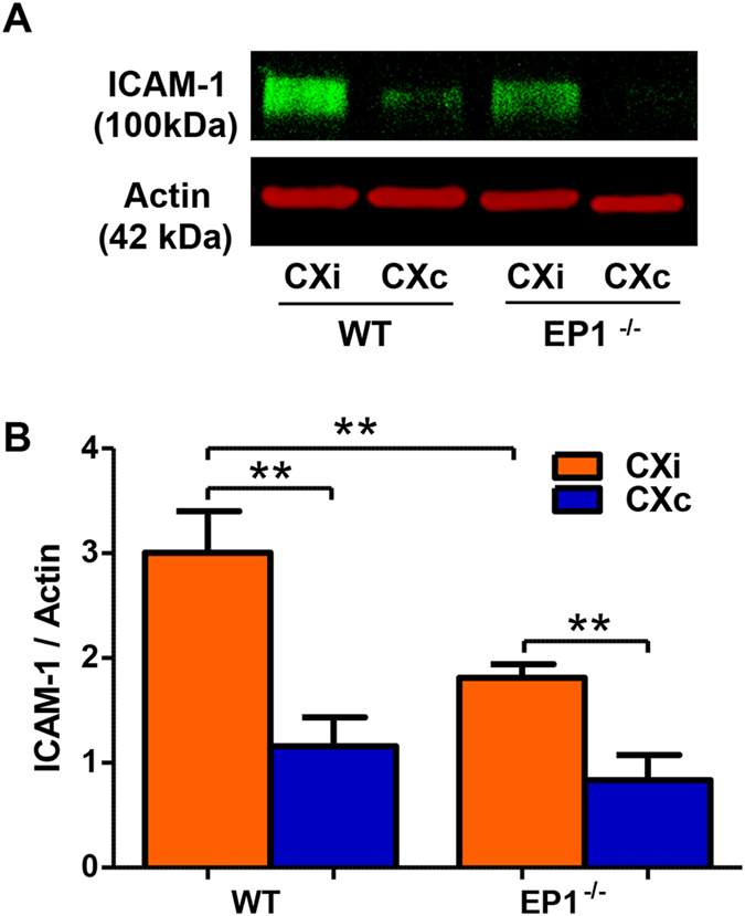
Figure 11. EP1−/− mice show reduced ICAM-1 levels in the ischemic cortex.

ICAM-1 levels were measured to determine whether EP1 deletion modulates ICAM-1 production in response to ischemia. (A) A representative Western blot for ICAM-1 in the mouse cortex. (B) Densitometric analysis of ICAM-1 levels showed an increase in the ischemic hemisphere compared to the contralateral hemisphere in both groups (P < 0.01, unpaired two-tailed t-test). ICAM-1 levels are significantly decreased in the ischemic EP1−/− mice compared to the WT (P < 0.01, unpaired two-tailed t-test). WT, N = 6; EP1−/−, N = 8. CXi = Cortex ipsilateral to stroke, CXc = Cortex contralateral to stroke.
