Fig. 2.

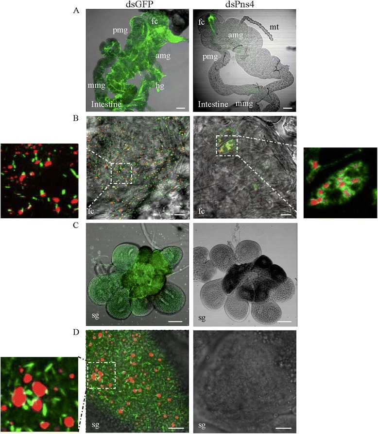
Microinjection of dsPns4 inhibited RDV infection and spread in insect vectors in vivo. At 12 days padp, the dissected intestines a and b and salivary glands (c and d) from leafhoppers receiving dsGFP or dsPns4 were immunolabeled with virus-FITC (green) (a and c), Pns4-FITC (green) (b and d) or Pns12-rodanmine (red) (b and d). The images with green fluorescence or red fluorescence were merged under a background of transmitted light. The enlarged images showing green fluorescence (Pns4-FITC) and red fluorescence (Pns12-rodanmine) of the merged images in the boxed areas in each panel, indicated that both of minitubules and diffusion of Pns4 distributed at the edge of viroplasms. fc, filter chamber; amg, anterior midgut; mmg, middle midgut; pmg, posterior midgut; hg, hindgut; mt, Malpighian tubules; sg, salivary gland. Bars, 100 μm (a and c) and 10 μm (b and d)
