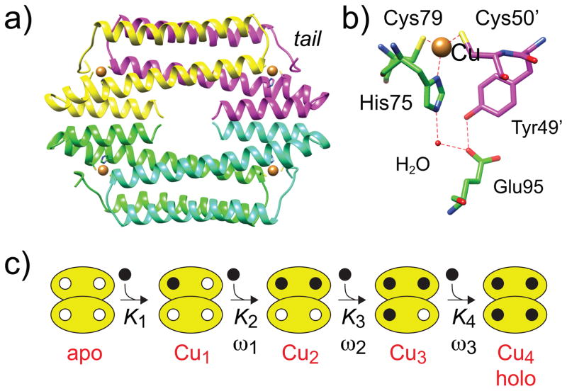
Figure 1.
Ribbon representation (a) of the structure of a thermophilic CsoR tetramer (PDB: 4M1P) closely related to B. subtilis CsoR studied here.[16] Each protomer is shaded differently, with the Cu(I) ions indicated by the brown spheres. The folded N-terminal tail (tail) is indicated. (b) Close-up of the Cu(I) binding pocket of CsoR. (c) Schematic representation of the step-wise Ki and ωi used this analysis.

