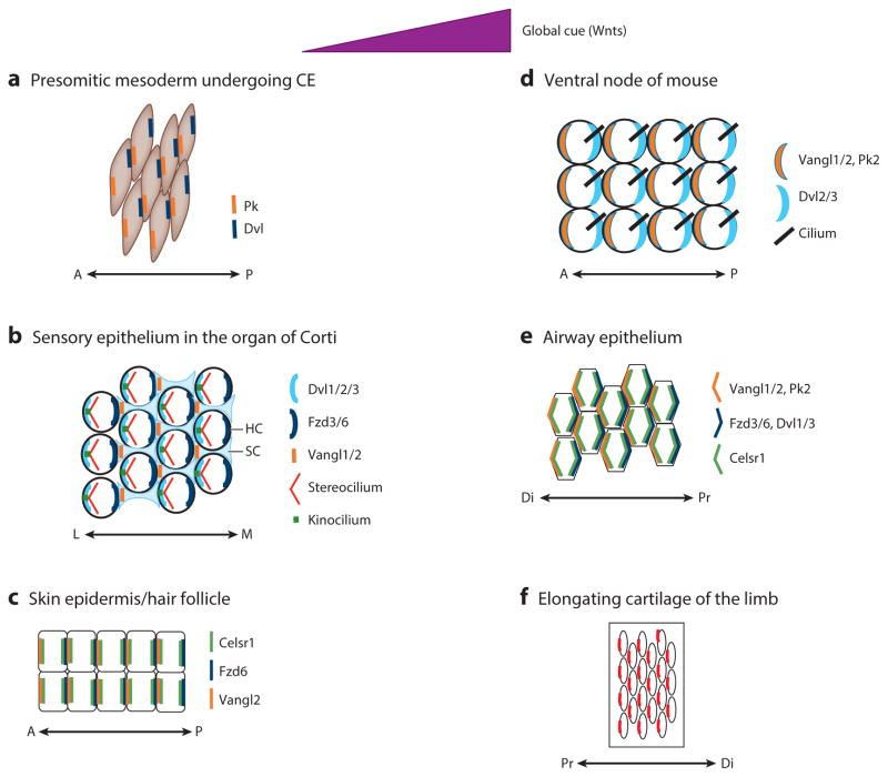
Figure 3.
Conserved asymmetrical localization patterns of core PCP components in six distinct epithelial and mesenchymal tissues (a–f ) in developing vertebrate embryos. In response to global cues (likely provided by Wnts), PCP is established. Molecularly, PCP establishment leads to coordinated, asymmetrical localization of core PCP components uniformly across all cells in any given tissue. It is interesting that the pattern of asymmetrical localization is conserved among different tissues. Moreover, the spatial relationships between global cue direction and the resulting asymmetrical pattern of polarity protein localization are also conserved. Abbreviations: A, anterior; CE, convergent extension; Di, distal; Dvl, Dishevelled; FzD, Frizzled; HC, hair cell; L, lateral; M, medial; P, posterior; PCP, planar cell polarity; Pk, Prickle; Pr, proximal; SC, supporting cell; Vangl, Van Gogh–like.

