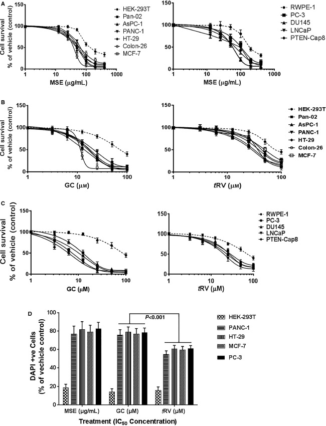
Figure 2.
Effect of MSE or GC on cancer cell proliferation and apoptosis. (a-c) MSE and GC inhibited proliferation of all human and mouse cancer cells in a concentration -dependent manner. GC significantly inhibited cancer cell proliferation compared with tRV. MSE or GC did not adversely affect normal cells. Data presented are means ± SD, and are representative of three independent experiments. (d) The bar graph shows the rate of apoptosis (percentage of apoptotic cells determined by DAPI staining) in the MSE and GC treatment groups, normalized to the vehicle (DMSO) treatment. DAPI-positive cells with characteristic nuclear condensation and DNA strand breaks for apoptosis were counted from 10 identical fields using a fluorescence microscope (Olympus) with × 40 magnifications. In contrast to the profound apoptosis induction in cancer cells, only marginal or very low levels of apoptosis were detected after normal cells were incubated with MSE or GC. A significant increase in apoptosis induction was observed after incubation with GC compared to tRV, p<0.001. The data are presented as the mean ± SD and are representative of three independent experiments.

