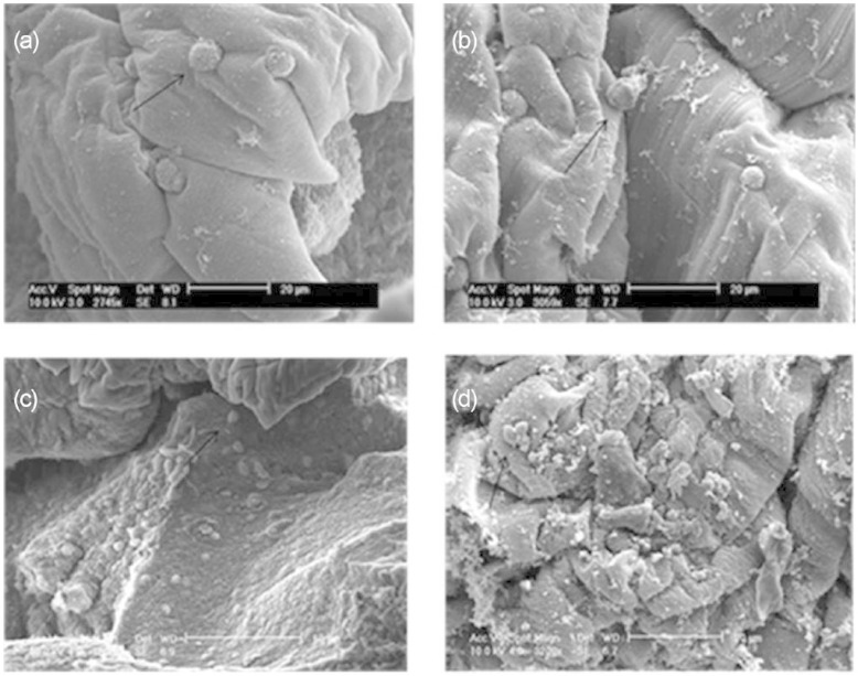
Figure 5.
Scanning electron micrographs of a cross-section of alginate disc showing human dermal fibroblasts encapsulated in alginate after 10 days in culture for (a) the control group in the absence of growth factors, (b) the treated group in the absence of growth factors, (c) the control group in the presence of growth factors, (d) the treated group in the presence of growth factors and (e) the alginate disc only. A blue dark region surrounding the cells shows the presence of GaG (arrow). Examples of viable cells are indicated with an arrow.

