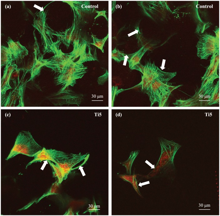
Figure 6.
Confocal laser scanning microscopy images of hMSCs growing on the (a, b) control microspheres and (c, d) Ti5 microspheres in ultra-low attachment plates at 7 days post culture. DMEM is used as the culture medium. Phalloidin stains the actin filaments of the cytoskeleton green, while propidium iodide stains the nuclei red. The white arrows in the images indicate the alignment of cytoskeletal filaments along the surface curvature.

