Figure 1.

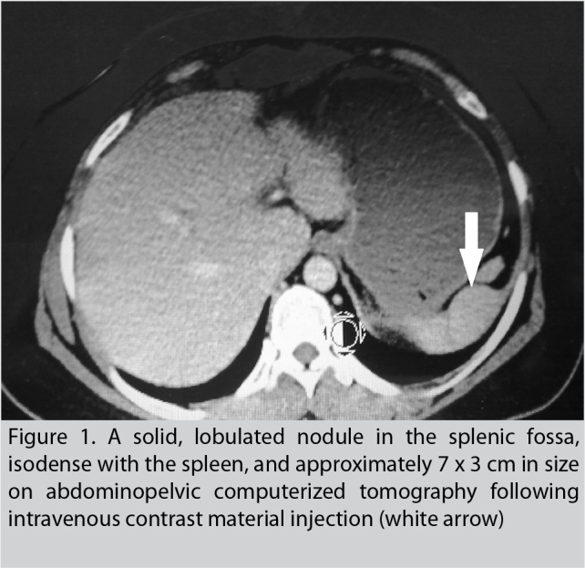
A solid, lobulated nodule in the splenic fossa, isodense with the spleen, and approximately 7 × 3 cm in size on abdominopelvic computerized tomography following intravenous contrast material injection (white arrow)

A solid, lobulated nodule in the splenic fossa, isodense with the spleen, and approximately 7 × 3 cm in size on abdominopelvic computerized tomography following intravenous contrast material injection (white arrow)