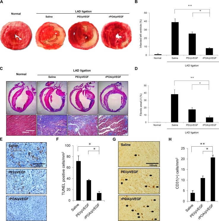
Fig 4. Histological analysis of gene therapy-treated MI hearts.
(A) Representative picture of myocardial sections stained with 2,3,5-triphenyltetrazolium chloride (TTC). Pale yellow indicates infarct region. (B) Ratio of infarcted to non-infarcted left ventricular myocardium (%) from LAD-ligated rats injected with saline, PEI/pVEGF, or rPOA/pVEGF (n = 5/group). *P < 0.0005, **P < 0.00001 vs. saline. (C) Representative myocardium sections stained with Masson’s trichrome (lower panel, 200×). Scale bar, 200 μm. (D) Percent of myocardial collagen fibrosis expressed as the ratio of fibrotic area to left ventricle area in LAD-ligated rats injected with saline, PEI/pVEGF, or rPOA/pVEGF (n = 5/group). *P < 0.005, **P < 0.00001. (E) Myocardial sections labeled with an antibody against CD31 to detect neovascularization 1 week after injection (400×). Scale bar, 100 μm. (F) Quantitative analysis of CD31-positive cells in ischemic myocardia treated with saline, PEI/pVEGF, or rPOA/pVEGF (n = 5/group). *P < 0.05, **P < 0.005.

