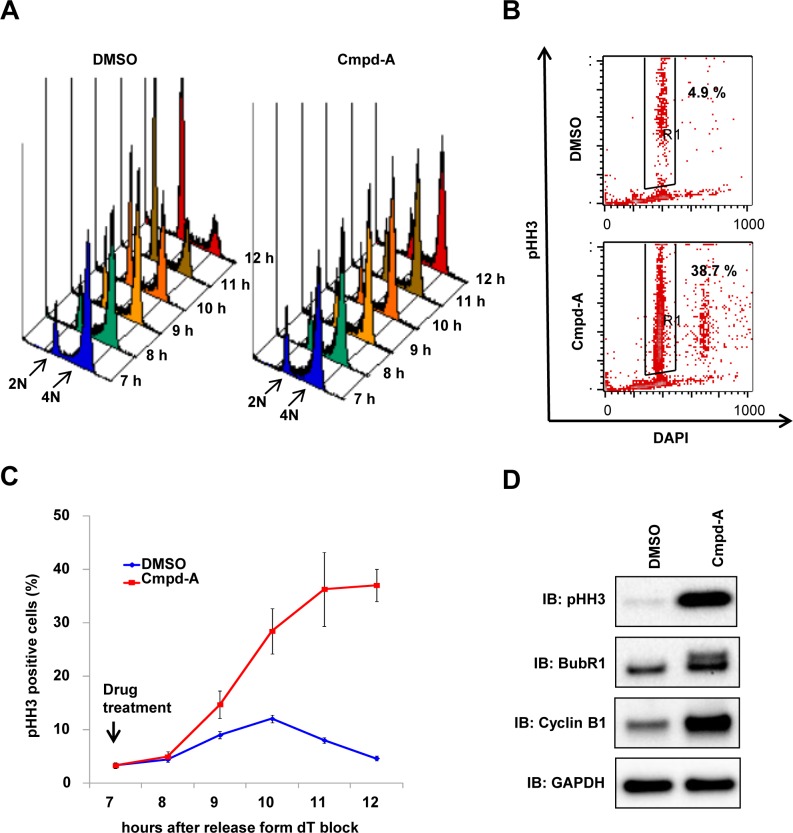
Fig 2. Cmpd-A induces prolonged mitotic arrest accompanied by SAC activation.
(A) Cell cycle histogram of synchronous HeLa cells treated with Cmpd-A (200 nM) or DMSO. Cmpd-A was added at the G2 phase (7 h after dT block release), and the cells were collected at the indicated time points for FACS analysis. (B) pHH3 in synchronous HeLa cells treated with Cmpd-A (200 nM) or DMSO. Cmpd-A was added at the G2 phase (7 h after dT block release), and the cells were collected 12 h after dT block release. Representative results are shown. (C) pHH3 elevation in synchronous HeLa cells treated with Cmpd-A (200 nM) or DMSO. Cmpd-A was added at the G2 phase (7 h after dT block release), and the cells were collected at the indicated time points for FACS analysis of pHH3 staining. The graph indicates quantified pHH3-positive cells (mean ± standard deviation; n = 3). Red and blue lines indicate Cmpd-A- and DMSO-treated HeLa cells, respectively. (D) Immunoblotting of mitosis markers in synchronous HeLa cells treated with Cmpd-A or DMSO. The cells were treated with Cmpd-A or DMSO as described in Fig 2A and collected 12 h after dT block release for immunoblotting.

