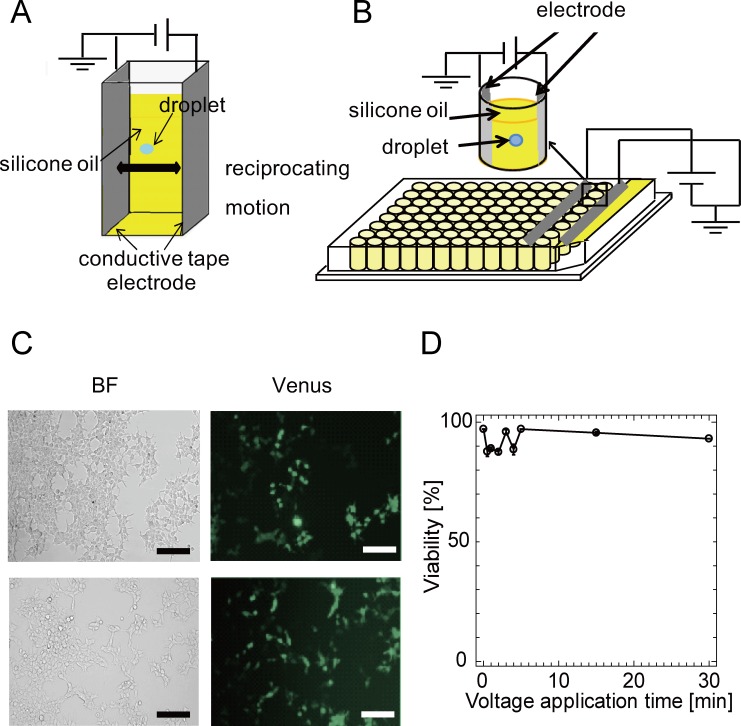
Fig 1. The droplet actuation device and cells transfected by the W/O droplet electroporation method.
(A), (B) Two pieces of conductive tape were set parallel on a single cuvette or 8 wells in a line for 96-well plastic microwell plates. The droplet continued to bounce between the edges of the two electrodes. One was the ground electrode, and the other was the high-voltage electrode. Images of droplets bouncing between the anode and cathode in each well are shown. Many cells in droplets can be transfected simultaneously. (C) The upper row : Bright-field (BF), fluorescence, and merge images of HEK293 cells 24 hours after transfection by W/O droplet electroporation. Cells were examined at 24 hours after transfection to evaluate the expression of fluorescent protein (Venus). Scale bars, 100 μm. The lower row : BF, fluorescence, and merge images of HEK293 cells 24 hours after transfection by lipofection. (D) Variation of cell viability with time of droplet actuation determined by trypan blue staining. All experiments were performed at least twice.

