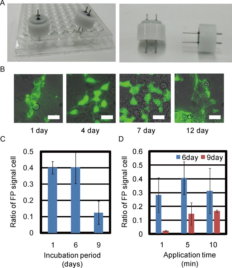
Fig 2. Screening of suitable conditions for W/O droplet electroporation.
(A) The pair of pin electrodes for single well of 96multi-well plate. (B) Merge image between bright-field (BF) and fluorescence images of HEK cells 1, 4, 7, and 12 days after W/O droplet electroporation. All scale bars, 30 μm. (C) HEK293 cells transfected with Venus fluorescent plasmid 1, 6, and 9 days after W/O droplet electroporation. Transfection efficiency was compared with 5-minute application time. (D) HEK293 cells transfected with Venus plasmid with application times of 1, 5, and 10 minutes 6 and 9 days after electroporation.

