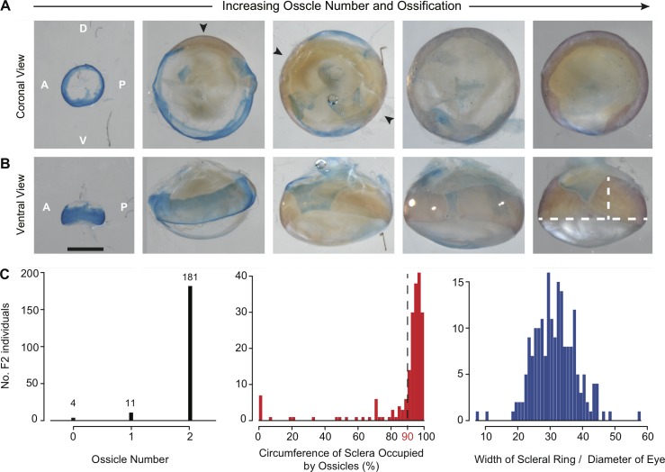
Fig 3. Limited phenotypic variation in scleral ossification among the progeny of a SF x CF F2 hybrid cross.
(A) Illustrative examples of reduced and normal scleral ossicle formation among 196 hybrid F2 progeny. Individual scleral ossicles are stained red with alizarin and highlighted with black arrows. (B) A ventral view of the same eyes as in (A). The length of the scleral ossicles varied dramatically, with some that occupy the entire circumference of the eye. White bars on the last image indicate where the width of the scleral ring and diameter of the eye were measured. (C) Distribution of the scleral phenotypes measured in this study. Both the number of scleral ossicles and the circumference of the sclera occupied by ossicles were highly skewed towards the wild-type phenotype of two ossicles that occupy >90% of the eye. Dotted line in the second panel indicates the threshold (90%) used to denote wild-type versus reduced scleral ossicles.

