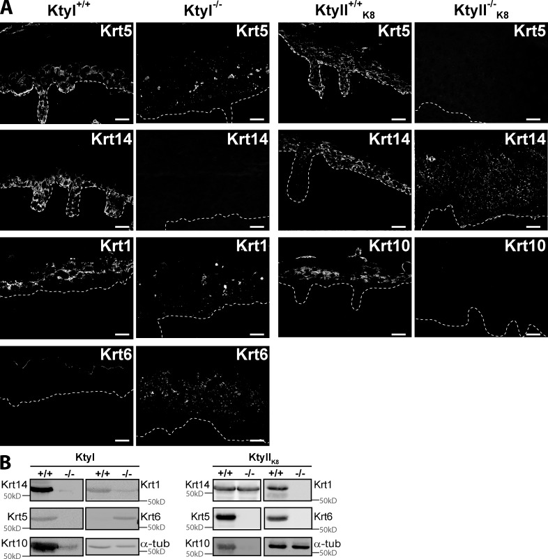
Figure 3.
Isotype-specific keratin expression and localization in KtyI−/− and KtyII−/−K8 epidermis. (A) Immunofluorescence analysis of keratin localization in WTs and KtyI−/− and KtyII−/−K8 epidermis at E18.5. Note absence of KIF in both KOs, suprabasal aggregates of Krt1 and Krt5 in KtyI−/−, and widespread aggregates of Krt14, but not of Krt10, in KtyII−/−K8. Basement membrane is indicated by white dotted lines. Bars, 20 µm. (B) Western blot analysis of Krt1, Krt5, Krt6, Krt10, Krt14, and α-tubulin as loading control in skin extracts of WT and keratin-deficient embryos at E18.5. Note positive correlation with A, showing a significant increase in Krt6 in KtyI−/− but unaltered presence of Krt14 in KtyII−/−K8 compared with KtyII+/+K8.

