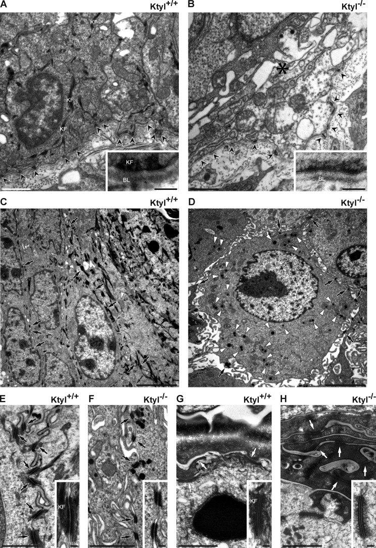
Figure 4.
Electron microscopy analysis of skin from KtyI+/+ and KtyI−/− E18.5 embryos depicting structural changes. (A and B) Hemidesmosomes (black arrowheads) of the stratum basale are readily detectable in KtyI−/− animals. Note, however, the absence of keratin filaments (KF) and occurrence of cytolysis in the basal cell layer of KtyI−/− epidermis (B, asterisk). Insets show higher magnification of hemidesmosomes together with the adjacent basal lamina (BL). (E and F) Desmosomes (black arrows) in the suprabasal layers of KtyI−/− epidermis are smaller and less frequent compared with KtyI+/+. Note the wide intercellular space in the stratum spinosum of KtyI−/− epidermis and intramitochondrial inclusions (white arrowheads). Insets in E and F show higher magnification of desmosomes. (G and H) Corneodesmosomes (white arrows) in the stratum corneum can be easily distinguished in KtyI−/− animals. Insets show higher magnification of corneodesmosomes. Bars: (insets) 100 nm; (A, B, and E–H) 1 µm; (C and D) 5 µm.

