Figure 5.
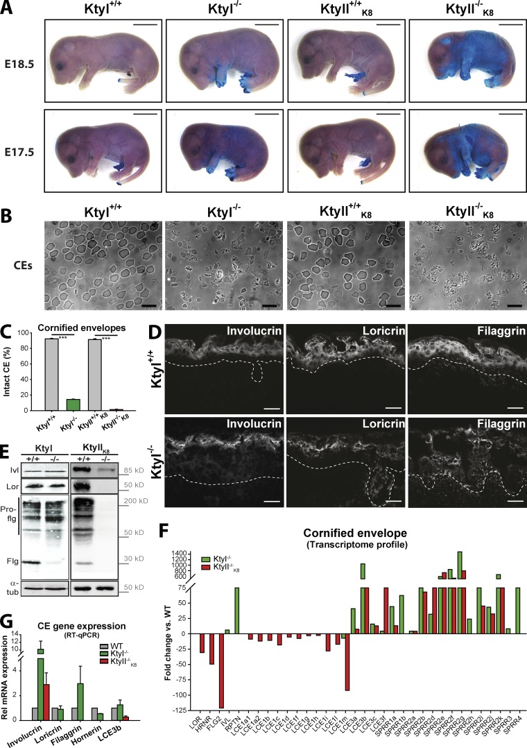
Distinct epidermal barrier defects of KtyI−/− and KtyII−/−K8 mouse embryos. (A) Toluidine blue assay indicating moderately delayed acquisition of dye-impermeable barrier in KtyI−/− and a severe delay in KtyII−/−K8 compared with WT embryos. Bars, 5 mm. (B and C) 85% of CEs from E18.5 embryos were disrupted in KtyI−/− compared with 99% in KtyII−/−K8 CE compared with WTs. Bars, 100 µm. (D) Immunofluorescence analysis of E18.5 epidermis revealing highly similar distribution of involucrin, loricrin, and filaggrin in KtyI+/+ and KtyI−/−. Note reduced filaggrin staining in KtyI−/− samples. The basement membrane is indicated by white dotted lines. Bars, 20 µm. (E) Western blotting of skin extracts from E18.5 embryos showing a decrease in involucrin (Ivl) and absence of loricrin (Lor) and filaggrin (Flg) in KtyII−/−K8, together with decreased processing of profilaggrin (Pro-flg, upper bands) into filaggrin (bottom bands) in KtyI−/− skin; α-tubulin (tub) served as loading control. (F) Transcriptome analysis exposing distinct changes in the expression of barrier protein-encoding genes in KtyI−/− and KtyII−/−. Note strong downregulation of a few barrier protein-encoding genes in KtyII−/−K8 only (in red); n = 3. (G) Quantitative RT-PCR analysis confirming the absence of transcription of loricrin, filaggrin, and hornerin genes in KtyII−/−K8 and upregulation of involucrin and loricrin mRNA in KtyI−/− skin. ***, P ≤ 0.001.

