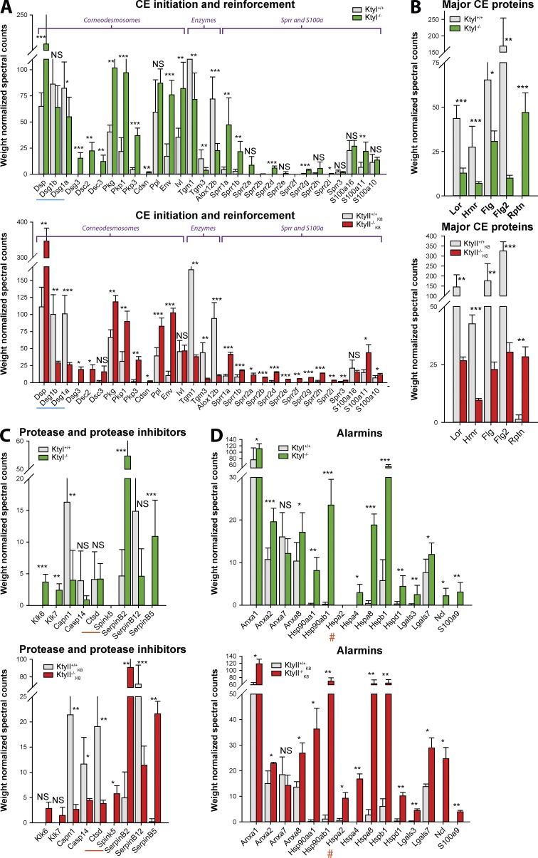
Figure 8.
CE proteome profile is keratin dependent. Grouping of CE constituents according to function in the presence and absence of type I and type II keratins. (A) CE initiation and reinforcement. Decrease in desmoglein-1 (underlined in blue), but increase in corneodesmosome and CE initiation and reinforcement proteins in KtyI−/− and KtyII−/−K8 compare d with WT. Less transglutaminase (tgm-1 and -3) and arachidonate 12-lipoxygenase (alox12b) are cross-linked in both KtyI−/− and KtyII−/−K8 compared with WT. (B) Except repetin, fewer major CE structural proteins are cross-linked in both KO strains compared with WT. (C) Cross-linking of proteases and protease inhibitors involved in terminal differentiation and barrier function is similarly changed in KtyI−/− and KtyII−/−K8 compared with WTs. Cathepsin-D (Ctsd) and Spink5 are underlined in orange and are only altered in KtyII−/−K8. (D) Enhanced cross-linking of alarmins in KtyI−/− and KtyII−/−K8 compared with WTs (red hash marks hspa2, which is upregulated in KtyII−/−K8 only). *, P ≤ 0.05; **, P ≤ 0.01; ***, P ≤ 0.001.

