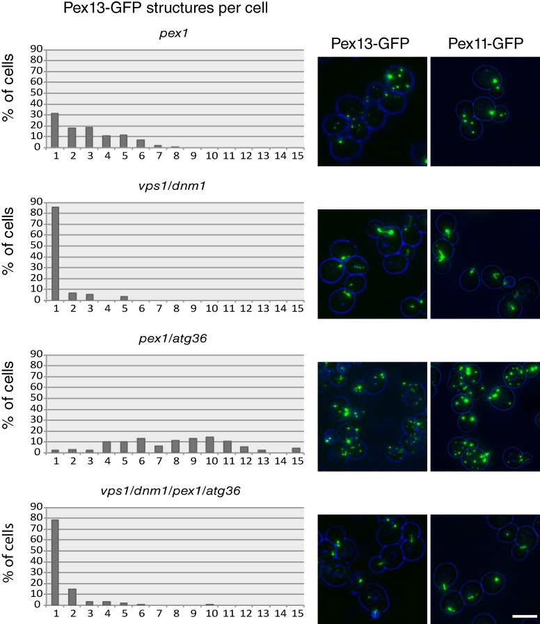
Figure 3.
Quantitation of membrane structures in pex1Δ, pex1Δ/atg3Δ6, vpsΔ1/dnm1Δ, and vps1Δ/dnm1Δ/pex1Δ/atg3Δ6 cells. Mutants expressing Pex11- or Pex13-GFP from their endogenous promoters (on plasmids) were imaged by epifluorescence microscopy. Cells were kept in a log phase on glucose-containing medium for 18 h before imaging. The doubling time of WT, vps1Δ/dnm1Δ, pex1Δ/atg36Δ, and vps1Δ/dnm1Δ/pex1Δ/atg36Δ strains under this condition was 101 min, 100 min, 106 min, and 115 min, respectively. The number of Pex13-GFP puncta per cell was determined for at least 200 cells per strain. Bar, 5 µm.

