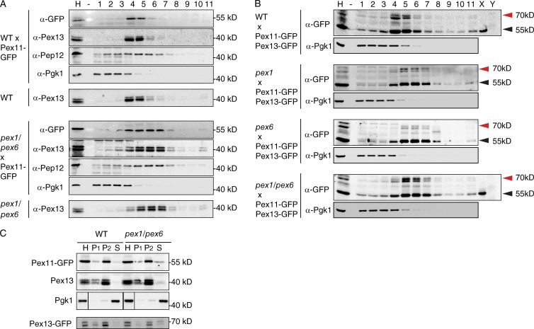
Figure 7.
Cofractionation of Pex11-GFP with Pex13 and Pex13-GFP in WT, pex1, pex6, and pex1/pex6 cells. (A and B) Homogenates (H; 800 g postnuclear supernatant) adjusted to 60% sucrose were separated by flotation analysis through sucrose equilibrium density gradient centrifugation. Fractions were collected from the bottom, and equal volumes were analyzed by Western blotting. Homogenates were prepared from glucose-grown cells of the strains as indicated. Cytosolic (Pgk1; A and B) and endosomal (Pep12; A) markers were included as the control for separation of membranes from cytosol. Pex13 is detected as a doublet, as Pex13 is prone to partial breakdown by proteolysis during subcellular fractionation (Elgersma et al., 1996). The samples in B were TCA precipitated and concentrated fourfold, as Pex13-GFP signals were weak. X and Y indicate control samples containing a P2 fraction of WT or pex1/pex6 cells expressing Pex11-GFP only and untransformed WT or pex1/pex6 cells. Red arrowheads indicate doublets of Pex13-GFP. Black arrowheads indicate Pex11-GFP. (C) Homogenates from WT and pex1/pex6 cells transformed with either Pex11-GFP or Pex13-GFP were separated by centrifugation first at 2,500 g and then at 25,000 g into a 2,500 g pellet (P1), a 25,000 g pellet (P2), and a 25,000 g supernatant fraction (S). Equivalent portions of each fraction were analyzed by Western blotting. Black lines indicate that intervening lanes have been spiced out.

