Figure 4.
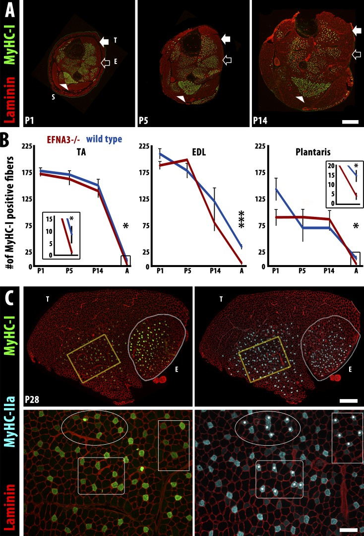
The loss of slow myofibers in ephrin-A3−/− mice occurs postnatally by conversion to IIA fast myofibers. (A) Immunohistochemistry showing laminin (red) and MyHC-I (green) expression in transverse cryosections of distal hindlimbs of ephrin-A3−/− mice at P1, P5, and P14. The soleus (S, arrowhead), TA (T, closed arrow), and EDL (E, open arrow) muscles can be identified by the spatial pattern of MyHC-I+ve myofibers in each section. Bar, 500 µm. (B) Slow myofiber number in TA, EDL, and plantaris of wild-type and ephrin-A3−/− (EFNA3−/−) muscles are not significantly different until after at least 2 wk of postnatal life (insets show slow myofiber number in adult TA and plantaris at a different scale). Error bars = SEM. *, P < 0.05; ***, P < 0.001. (C) MyHC-I+ve myofibers in ephrin-A3−/− muscle sections at P28 (green, left) frequently coexpress MyHC-IIa (cyan, right), indicating that they are in transition to a faster fiber type; laminin (red) outlines myofibers on both sides. Insets (yellow boxes) are magnified under each panel: three regions are identified in each inset to facilitate comparison, and asterisks indicate MyHC-IIa+ve myofibers that are also MyHC-I+ve. Bars: (montages) 500 µm; (insets) 100 µm.

