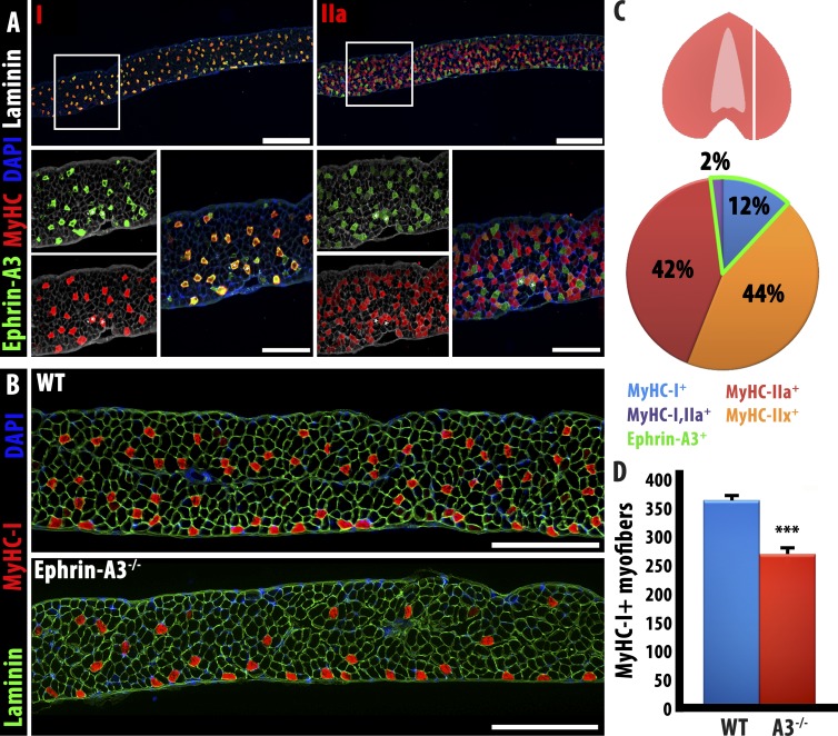
Figure 6.
Ephrin-A3−/− diaphragm muscle displays a phenotype intermediate between TA and soleus muscles. (A) As noted in hindlimb muscles, ephrin-A3 expression is detected on all MyHC-I+ve myofibers in the diaphragm; in sections taken as indicated in the cartoon at right, MyHC-I is expressed by 14% of myofibers scored, all of which also express ephrin-A3. Bars: (top) 400 µm; (bottom) 100 µm. (B) Equivalent sections of wild-type and ephrin-A3−/− adult diaphragm show a reduction, but not loss, of slow myofibers. Bars, 500 μm. (C) Representation of the plane of section in A and B and quantification of muscle fiber types and ephrin-A3 expression in WT diaphragm. (D) Quantification of Type I fibers through the same plane of section in wild-type and ephrin-A3−/− adult diaphragm; error bars represent SEM. WT, wild type. ***, P < 0.001.

