Figure 3.
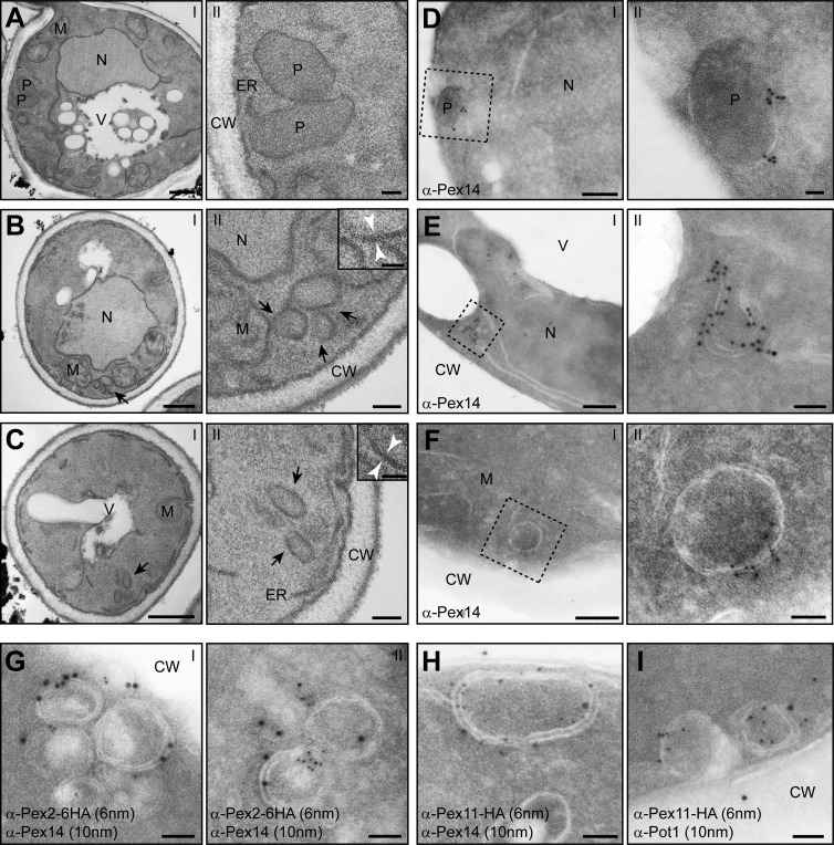
pex1 and pex6 cells harbor large peroxisomal ghosts that contain Pex2, Pex11, and Pex14 but lack thiolase. (A–C) EM analysis of ultrathin sections of KMnO4-fixed BY4742 WT (A, I and II), pex1 (B, I and II), or pex6 cells (C, I and II). Cells were grown for 16 h in the presence of oleic acid. The insets in panel II of B and C show higher magnifications, which reveal the double membranes in cross sections. (D–F) Immunolabeling of cryosections of aldehyde-fixed, oleic acid–grown cells labeled with α-Pex14 antibodies (WT: D, I and II; pex1: E, I and II; pex6: F, I and II). (G and H) Ultrathin sections of aldehyde-fixed, oleic acid–grown pex1 cells, which produce C-terminal HA-tagged Pex2 (G) or Pex11 (H), were labeled using rabbit α-Pex14 and mouse α-HA antibodies and detected using α-rabbit 10-nm and α-mouse 6-nm gold particles. (I) Double immunolabeling using α-thiolase antibodies (Pot1), showing that the Pex11-labeled structures do not accumulate thiolase. CW, cell wall; M, mitochondrion; N, nucleus; P, peroxisome; V, vacuole. Bars: (A–C, I) 500 nm; (D–F, I) 200 nm; (A–C, II) 100 nm; (B, II, inset; C, II, inset; D, II; E, II; F, II; and G–I) 50 nm.

