
Figure 2.
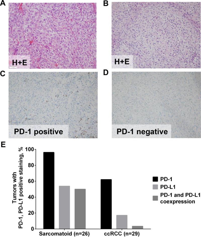
Representative Immunohistochemical Staining of PD-1 Expression in RCC With Sarcomatoid Features and Clear Cell RCC. Staining of sarcomatoid (A and C) and ccRCC (B and D) samples. A and B, Hematoxylin-eosin staining (H+E). C and D, Staining with anti–PD-1 antibody showing positive staining of sarcomatoid RCC (C) and negative staining of ccRCC (D). All panels, original magnification ×20. E, Percentage of tumors with PD-L1 expression (clone 130021), PD-1+ tumor-infiltrating lymphocytes, or coexpression of PD-L1 and PD-1 in sarcomatoid RCC or ccRCC.