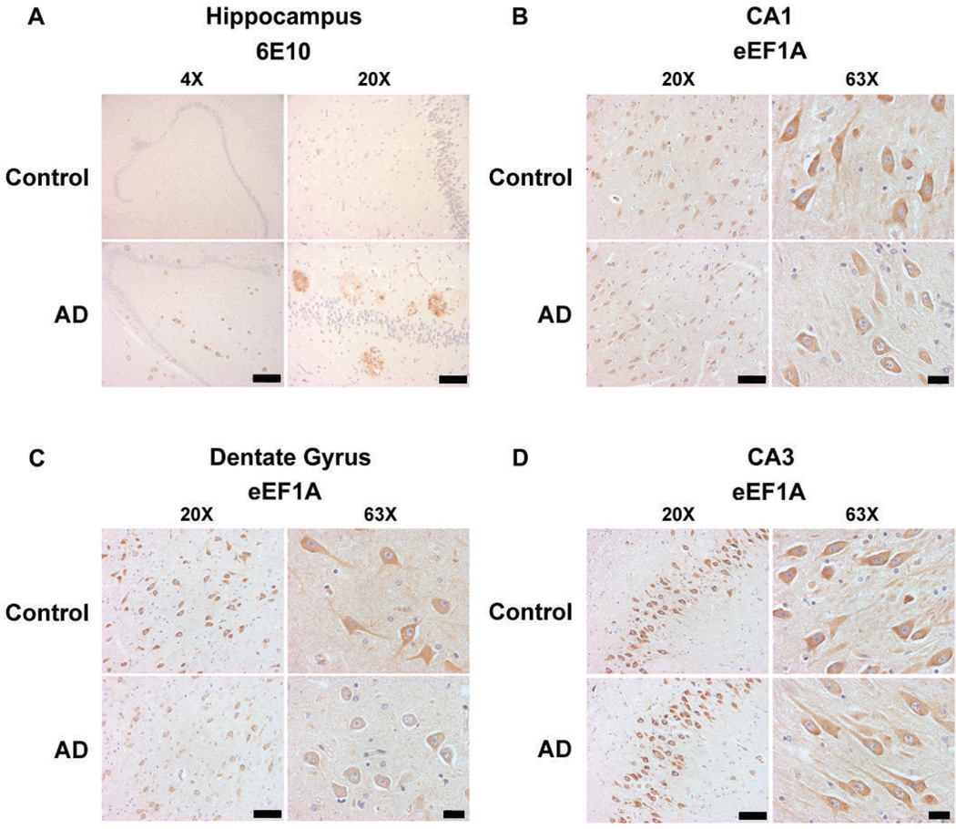
Figure 2. Hippocampal neuronal eEF1A levels are reduced in human AD patients.
(A) Immunohistochemistry with Aβ antibody (6E10) demonstrates abundant hippocampal “plaques” involving hilus, CA fields, and dentate gyrus of AD patients (AD), whereas controls (CT) have very little plaque deposition. Representative images are shown for 4× (scale bar 1 mm) and 20× (scale bar 200 µm) magnification. (B) Compared with controls, patients with AD have decreased eEF1A immunostaining in CA1 pyramidal neurons. (C) In hilar neurons (CA4) of the dentate gyrus, AD patients also displaya pronounced reduction in eEF1A levels. (D) Levels of eEF1A immunostaining in CA3 pyramidal neurons in AD patients are not different from controls. For (B), (C), and (D), representative images are shown for 20× (scale bar 200 µm) and 63× (scale bar 50 µm) magnification. Images shown represent results from four independent experiments.

